
Heart World Conference 2026
Scientific Committee
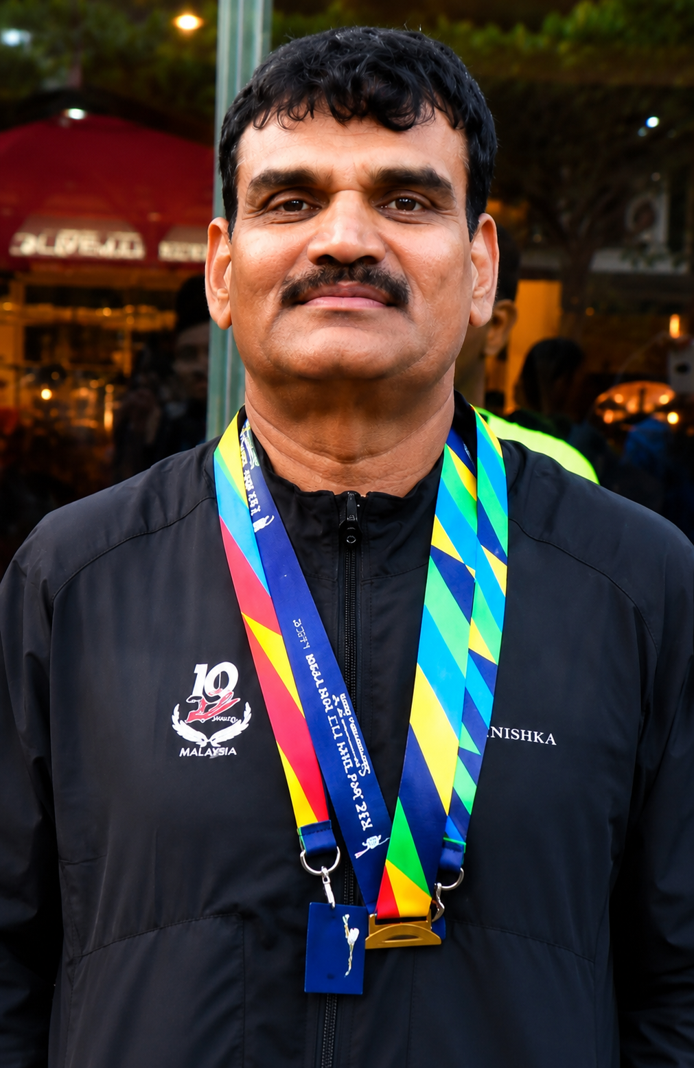
Anand Balaram Patil
Anand Balaram Patil
- Designation: Department of Clinical Cardiology and Cardiac Rehabilitation, Medicover Hospitals, Navi Mumbai, Maharashtra,
- Country: India
Biography
Dr. Anand Balaram Patil, MBBS, MS, FACC, CCRP, is Lead Coordinator of Cardiac Rehabilitation and Preventive Cardiology at Medicover Hospitals, Navi Mumbai, India, and Founder–Director of the Dr. ABP Research Institute for Fitness & Rehabilitation. A clinician-scientist and exercise physiologist, his research focuses on precision cardiac rehabilitation, cardiopulmonary exercise testing (CPET), and exercise-based management of heart failure. He developed the Patil Protocol, integrating the Qualify Quantif Recovery Framework (QQRF) and High-Volume Restricted-Intensity Training (HVRIT) to personalise rehabilitation in ischaemic HFrEF. Dr. Patil is a Fellow of the American College of Cardiology (FACC) and has presented his research at leading international cardiovascular scientific meetings.
Abstract
Exercise-based cardiac rehabilitation is a Class I recommendation for patients with heart failure with reduced ejection fraction (HFrEF). However, evidence regarding prolonged high-volume aerobic training performed at restricted physiological intensity in patients with ischemic HFrEF, previous ventricular tachyarrhythmia, and implantable cardioverter-defibrillator (ICD) therapy remains extremely limited. Conventional cardiac rehabilitation generally prioritizes moderate exercise doses and symptom-limited progression. The unresolved clinical dilemma is whether high training volume can be accumulated predominantly below an individualised physiological ceiling in a carefully selected, experienced endurance athlete with stable HFrEF and an ICD, without triggering malignant arrhythmias or worsening ventricular dysfunction. We hypothesized that implementing the Patil Protocol via the Qualify-Quantify-Recovery Framework (QQRF) enables safe accumulation of high-volume endurance exercise, resulting in positive left ventricular reverse remodelling and functional enhancement without increasing arrhythmic risk.
Methods
Patient Population: A 61-year-old South Asian male recreational endurance athlete presenting with acute anterior STEMI, LAD stent thrombosis, cardioembolic stroke, VT/VF
arrest, and secondary-prevention dual-chamber ICD implantation, resulting in ischaemic HFrEF (baseline LVEF 32%).
Exercise Intervention: A four-phase, CPET-calibrated hybrid rehabilitation programme utilising High-Volume Restricted-Intensity Training (HVRIT) and High-Volume Safety-Interval Training (HVSIT). Routine training targeted an individualised aerobic ceiling of <130 beats/min, progressing long-session capacity from approximately 2 hours up to 10 hours in selected sessions.
CPET: Symptom-limited cardiopulmonary exercise testing performed at baseline and longitudinally to establish ventilatory thresholds and guide individualised exercise prescription.
Echo: Serial comprehensive echocardiography to assess left ventricular ejection fraction (LVEF) and left ventricular end-diastolic volume (LVEDV).
Biomarkers: Serial assessment of B-type natriuretic peptide (BNP) and comprehensive evaluation of renal stability (serum creatinine and eGFR).
Statistics: Due to the single-patient N-of-1 longitudinal design without replicate independent observations, descriptive changes are reported without inferential p-values.
Results
Peak VO‚ : Formal CPET peak oxygen uptake improved from 14.2 to 19.1 mL/kg/min (+35%).
EF: Left ventricular ejection fraction (LVEF) improved from 32% to 42% (+31% relative change).
LV Volumes: Left ventricular end-diastolic volume (LVEDV) decreased from 156 mL to 124 mL (-20%), indicating favourable reverse remodelling.
BNP: B-type natriuretic peptide levels decreased substantially from 312 pg/mL to 89 pg/mL (-71%), reflecting reduced neurohormonal activation.
NYHA: Functional class improved from NYHA Class III to Class I–II.
HR Recovery: Demonstrated favourable autonomic adaptation with more stable post-exercise heart rate recovery trends.
VE/VCO‚ : Ventilatory efficiency improved alongside enhanced cardiopulmonary coupling.
Additional Outcomes: Stroke volume increased from 48 to 60 mL, resting cardiac output improved from 3.8 to 5.1 L/min, and renal function remained stable (creatinine 1.0 to 1.1 mg/dL; eGFR 79 to 76 mL/min/1.73 m²). During Ironman 70.3 Tallinn, the patient experienced acute respiratory distress compatible with swimming-induced pulmonary oedema (SIPE) leading to a DNF, serving as an important safety boundary marker. No appropriate ICD therapies or heart-failure rehospitalisations were documented during the 20-month follow-up.
Conclusion
Clinical Implications: This case demonstrates the clinical feasibility of a closely monitored, high-volume but restricted-intensity rehabilitation strategy in a highly selected, experienced heart failure patient. It highlights that exercise volume and intensity can be successfully decoupled. However, the SIPE event underscores the critical distinction between controlled terrestrial training and high-demand open-water competitive exposure.
Future Multicentre Validation: While hypothesis-generating for precision cardiac rehabilitation, these findings require prospective, multicentre validation across broader HFrEF populations before wider clinical generalisation.
Keywords: Heart failure with reduced ejection fraction; Cardiac rehabilitation; Cardiopulmonary exercise testing; High-Volume Restricted-Intensity Training; Patil Protocol; Reverse remodelling; Implantable cardioverter-defibrillator; Exercise physiology.
Welcome to the Heart World Conference 2026, a premier international conference committed to discussing the entire gamut of heart science, technolog ...
Read More>>
Organizer
